Upper Ankle Diagram. Foot drop can result if there is injury to the dorsiflexors or to any point along the neural. They serve as shock asborbing structures that support body weight and distribute stress evenly during walking.
Grade of aluminum. The base of this pyramid opens anteriorly onto the face while the apex is pointed posteromedially towards the center of the skull. Most first year veterinary students have a misconception of the term leg anatomically the term leg means the part of the hind limb that extends from the stiffle joint to the hock joint knee to ankle or tibia and fibula bones region.
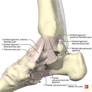
It is formed by the bones of the leg tibia and fibula and the foot talus.
The tibia the fibula and the talus. The medial collateral or deltoid ligament and lateral collateral ligament. Maxilla zygomatic bone lacrimal bone palatine bone frontal bone ethmoid bone and sphenoid bone. Its articulating surfaces ligaments movements and clinical.