Ekg Diagram Explanation. Schematic diagram of normal sinus rhythm for a human heart as seen on ecg the first upward pulse of the ekg signal the p wave is formed when the atria the two upper chambers of the heart contract to pump blood into the ventricles. By placing these electrodes at appropriate parts of body.
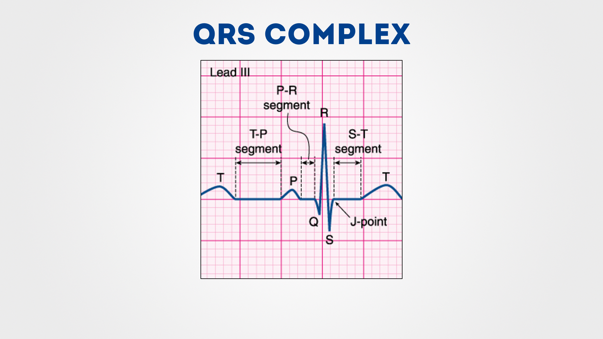
Septal view of the heart. In sinus rhythm there should be a p wave preceding each qrs complex. The other end of electrode passes through defibrillator protection circuit.
Figure 1 ekg tracing step 1 rate the first step is to determine the rate which can be eyeballed by the following technique.
Sensors attached to the skin are used to detect the electrical signals produced by your heart each time it beats. P waves represent atrial depolarization. Sensors attached to the skin are used to detect the electrical signals produced by your heart each time it beats. Schematic diagram of normal sinus rhythm for a human heart as seen on ecg the first upward pulse of the ekg signal the p wave is formed when the atria the two upper chambers of the heart contract to pump blood into the ventricles.